Optical Illusions
Optical Illusions can use color, light and patterns to create images that can be deceptive or misleading to our brains. The information gathered by the eye is processed by the brain, creating a perception that in reality, does not match the true image. Perception refers to the interpretation of what we take in through our eyes. Optical illusions occur because our brain is trying to interpret what we see and make sense of the world around us. Optical illusions simply trick our brains into seeing things which may or may not be real.
The Blind Spot
One of the most dramatic experiments to perform is the demonstration of the blind spot. The blind spot is the area on the retina without receptors that respond to light. Therefore an image that falls on this region will NOT be seen. It is in this region that the optic nerve exits the eye on its way to the brain. To find your blind spot, look at the image below or draw it on a piece of paper:

To draw the blind spot tester on a piece of paper, make a small dot on the left side separated by about 6-8 inches from a small + on the right side. Close your right eye. Hold the image (or place your head from the computer monitor) about 20 inches away. With your left eye, look at the +. Slowly bring the image (or move your head) closer while looking at the +. At a certain distance, the dot will disappear from sight…this is when the dot falls on the blind spot of your retina. Reverse the process. Close your left eye and look at the dot with your right eye. Move the image slowly closer to you and the + should disappear.
Depth Perception
Two eyes are better than one, especially when it comes to depth perception. Depth perception is the ability to judge objects that are nearer or farther than others. To demonstrate the difference of using one vs. two eyes to judge depth hold the ends a pencil, one in each hand. Hold them either vertically or horizontally facing each other at arms-length from your body. With one eye closed, try to touch the end of the pencils together. Now try with two eyes: it should be much easier. This is because each eye looks at the image from a different angle. This experiment can also be done with your fingers, but pencils make the effect a bit more dramatic.
Visual Illusions
What you see is not always what is there. Or is it? The eye can play tricks on the brain. Here are several illusions that demonstrate this point.
The Magic Cube
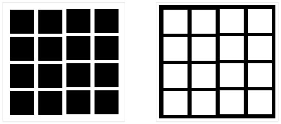
Stare at the middle of the picture with black squares between 15 to 30 seconds. Are those really dots that appear at the corners of the squares? What happens if you focus on a dot? Now look at the middle of the picture with the white squares. Do you see the dots again? What color are they this time?

X-Ray Vision
Do you have “X-Ray Vision?” You may be able to see through your own hand with this simple illusion. Roll up a piece of notebook paper into a tube. The diameter of the tube should be about 0.5 inch. Hold up your left hand in front of you. Hold the tube right next to the bottom of your left “pointer” finger in between you thumb.
Look through the tube with your RIGHT eye AND keep your left eye open too. What you should see is a hole in your left hand!! Why? Because your brain is getting two different images…one of the hole in the paper and one of your left hand.
After Images
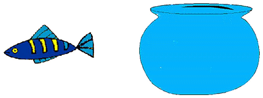
Here’s an example of creating an afterimage. Can you put the fish in the bowl? Try this. Stare at the yellow stripe in the middle of the fish in the picture below for about 15-30 seconds. Then move your gaze to the fish bowl. You should see a fish of a different color in the bowl. It helps if you keep your head still and blink once or twice after you move your eyes to the bowl. The afterimage will last about five seconds.

What’s Happening? In the retina of your eyes, there are three types of color receptors (cones) that are most sensitive to either red, blue or green. When you stare at a particular color for too long, these receptors get “tired” or “fatigued.” When you then look at a different background, the receptors that are tired do not work as well. Therefore, the information from all of the different color receptors is not in balance. Therefore, you see the color “afterimages.”
Baby’s First Eye Exam
A baby’s first visit to an optometrist should happen between 6 and 12 months of age. This is a critical time for eye and vision development and early detection of any problems can help with long term vision.
During the eye exam, the infant will sit on his or her parent’s lap. Dr. Curtis uses different lights, toys, and lenses to check that the infant’s eyes are working together and that there are no significant issues that will interfere with proper vision development. Specially formulated eye-drops are used to dilate the infant’s pupils which afford Dr. Curtis a better look inside the eye to ensure good health. Infants generally find the eye exam painless and fun!
Did you know that 1 out of 10 children is at risk from undiagnosed vision problems, and 56% of mothers and expectant mothers are not certain of when the most critical age is for the development of eyesight.


